
Introduction
Pre-eclampsia has been recognized for more than two thousand years, but it is still one of the most puzzling obstetrical syndromes. Its symptoms and causes are varied. Traditionally, doctors have used high blood pressure and protein in the urine as the main signs for diagnosis. However, new research shows that high blood pressure is a visible sign of deeper problems in the small blood vessels, which often start before any change in blood pressure is noticed.
This leads to an important question: are we treating a widespread blood vessel problem, or are we just observing a breakdown in communication between the placenta and the mother’s blood vessels?
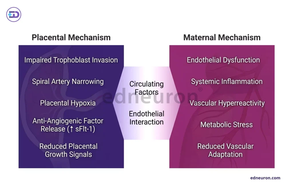
Fig 1: Dual-panel depiction of pre-eclampsia highlighting divergent placental insufficiency and maternal systemic dysfunction pathways.
.
The Mechanistic Divergence: Placental vs. Maternal Phenotypes
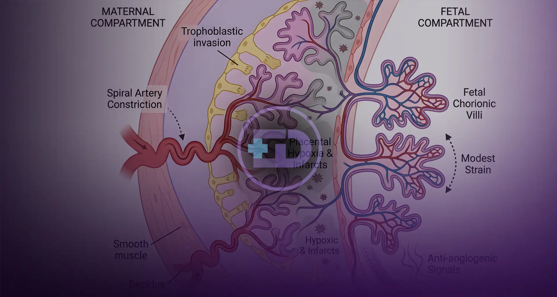
One of the main difficulties in treating pre-eclampsia is that it is not a single disease, but rather has different forms. It is usually divided into early-onset (before 34 weeks) and late-onset (34 weeks or later) types. Early-onset pre-eclampsia is often seen as a problem with the placenta, starting when certain cells fail to change the blood vessels as they should in the first trimester. As a result, the arteries retain their thick, muscular walls, leading to rapid, forceful blood flow that can damage the placenta’s fragile tissues. This stress triggers the first stage of the disease, characterized by damage to the outer layer of the placenta and the release of harmful substances.
On the other hand, late-onset pre-eclampsia often happens in women whose placentas look normal. This suggests that the problem may be the mother’s body not adjusting well to the changes of late pregnancy. Factors like obesity, long-term high blood pressure, and older age can make the mother’s blood vessels less able to handle the normal aging of the placenta. Even though early and late-onset pre-eclampsia start differently, they both end up causing a state in the body where blood vessels become very sensitive to certain hormones.
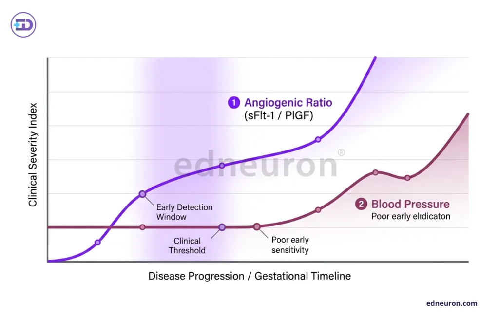
Fig 2: Early rise of angiogenic imbalance precedes and outperforms blood pressure in reflecting disease severity in pre-eclampsia.
The Angiogenic Dilemma: Ratios vs. Sphygmomanometry
Using blood pressure readings of 140/90 mmHg or higher to diagnose pre-eclampsia is now seen as too simple and possibly risky. Studies show that examining the balance of certain blood proteins, especially the sFlt-1 to PlGF ratio, provides a clearer picture of what is happening in the placenta than simply measuring blood pressure. The level of sFlt-1, which blocks the effects of important growth factors, often increases up to six weeks before any symptoms appear.
This difference in diagnosis creates a real challenge for doctors. A woman may seem healthy, but if her sFlt-1/PlGF ratio is above 66, she is at high risk of developing pre-eclampsia soon. On the other hand, if these markers are normal, doctors can be almost certain for a week that she does not have pre-eclampsia, which helps avoid unnecessary hospital stays for women with high blood pressure but no real disease. The real success in care comes from understanding these underlying changes, not just measuring blood pressure.
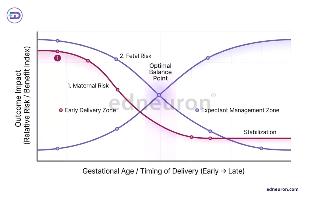
Fig 3: Opposing maternal and fetal risk curves illustrate the optimal timing of delivery in pre-eclampsia.
Clinical Outcomes and the Delivery Paradox
The only true cure for pre-eclampsia is delivering the placenta, but this solution brings its own risks. Doctors must weigh the mother’s risk of serious complications like brain swelling or HELLP syndrome against the baby’s risk from being born too early. Treatments like steroids to help the baby’s lungs and magnesium sulfate to prevent seizures are common, but they do not fix the damage to the mother’s blood vessels.
Furthermore, the resolution of postpartum symptoms does not signify a return to baseline health. Pre-eclampsia is now recognized as a potent “stress test” for future cardiovascular disease, with affected women facing a nearly fourfold increased risk of chronic hypertension and a doubled risk of stroke later in life. This suggests that the maternal endothelium may remain “primed” for vascular injury long after the offending placenta is removed.
Conclusion
Understanding pre-eclampsia as a complex problem involving blood vessel growth, not just high blood pressure, means doctors need to change how they monitor patients. Instead of just monitoring blood pressure, they should focus on the health of small blood vessels. Since there is no medicine that cures pre-eclampsia, the best approach is to realize that normal blood pressure can hide ongoing damage to the mother’s blood vessels.
References
- Rolnik DL, Wright D, Poon LC, O’Gorman N, Syngelaki A, de Paco Matallana C, et al. Prediction and prevention of late-onset pre-eclampsia. Front Med (Lausanne). 2024;11:1459289.
- Uzan J, Carbonnel M, Piconne O, Asmar R, Ayoubi JM. Pre-eclampsia: pathophysiology, diagnosis, and management. Vasc Health Risk Manag. 2011;7:467–74.
- Rana S, Lemoine E, Granger JP, Karumanchi SA. Pre-eclampsia: pathogenesis, novel diagnostics and therapies. Circ Res. 2019;124(7):1094–112.
- Staff AC, Benton SJ, von Dadelszen P, Roberts JM, Taylor RN, Powers RW, et al. Redefining pre-eclampsia using placenta-derived biomarkers. BMJ. 2019;366:l2381.
- Singh A, Kumar R, Sharma S. Etiopathology and outcome in pre-eclampsia. Asian J Health Sci. 2024;10(2):1–8.
- Kenny LC, Broadhurst DI, Dunn W, Brown M, North RA, McCowan LME, et al. Robust early pregnancy prediction of later pre-eclampsia using metabolomic biomarkers. Placenta. 2010;31(9):778–82.

Author: A D
A surgically inclined medical trainee with a growing interest in brain, spine, and cardiothoracic systems, aspiring to build a career in advanced operative care. He aims to become a patient-centered surgeon who integrates evolving medical technologies with precise clinical judgment. With a strong foundation in surgical sciences, he has gained clinical exposure through case postings involving breast pathologies, chronic ulcers, and diabetic foot, along with observational experience in hernia repair, appendectomy, and cholecystectomy. Academically, he has assisted research work on breast cancer and developed a case report on a complicated hernia surgery, alongside contributing multiple review articles across disciplines. He is currently engaged in ongoing research exploring the role of nutrition in health and disease. MBBS (MS4) GMC Nagpur, India