

Clinical Vignette
A 58-year-old woman presents with ten days of ascending leg weakness, diminished reflexes at the ankles, and a vague history of a respiratory illness three weeks prior. The clinical team plausibly suspects Guillain–Barré syndrome. When a lumbar puncture was performed, the total CSF protein was 72 mg/dL, and the white cell count was 2 per microlitre.
Albuminocytologic dissociation
The team nods in recognition – a pattern they predicted, a finding that feels confirmatory. Two weeks later, the patient’s weakness progressed despite intravenous immunoglobulin. An MRI of the thoracic spine, ordered almost as a safety net, revealed an epidural mass compressing the cord at T6. She had small-cell lung carcinoma.
The Origins
In 1916, Guillain-Barré and Stohl described a syndrome of acute flaccid paralysis accompanied by elevated CSF protein without a cellular reaction, a phenomenon that became eponymous and, in the decades that followed, iconic. Albuminocytologic dissociation, or ACD, became neurological shorthand for immune-mediated polyradiculoneuropathy in a way that no other CSF finding has managed so far. The problem is not that the association is wrong, but that it has become reflexive. Neurology has a long tradition of pattern recognition, and pattern recognition is not without virtue. But patterns derive their clinical utility from the probability with which they operate from prevalence, from pretest likelihood, and from the diagnostic weight of surrounding evidence. Extracted from those, a pattern becomes a shortcut, and shortcuts, as this case illustrates, can close diagnostic inquiry at precisely the moment it should open further.
Conceptual Interpretation
Albuminocytologic dissociation is a physiological clue – an archetype of altered neurochemical dynamics across the blood–nerve or blood–CSF interface.
To understand why ACD is a nonspecific finding, one must understand what it actually measures.
The cerebrospinal fluid is not just a passive filtrate of plasma. It is maintained by active transport mechanisms at the choroid plexus and robustly regulated permeability at the blood–CSF and blood–nervebarriers. CSF protein rises when these barriers are disrupted, allowing albumin, which is normally excluded from the intrathecal compartment, to leak across damaged or inflamed neural tissue. Importantly, this barrier dysfunction need not reflect lymphocyte trafficking. In Guillain–Barré syndrome and its variants, the mechanism is immune-mediated inflammation of nerve roots and peripheral nerves. An immune attack by anti-ganglioside autoantibodies at the level of the spinal nerve roots disrupts the blood–nerve barrier locally, allowing unbridled entry of albumin in the surrounding CSF. In chronic inflammatory demyelinating polyneuropathy (CIDP), where segmental demyelination and remyelination occur, a slower but analogous process operates. Similarly, the same final pathway, i.e., barrier dysfunction, impaired protein clearance, intrathecal accumulation, can be triggered by entirely different forces: mechanical compression of the spinal cord or cauda equina, obstruction of CSF flow by a mass or adhesion, infiltration of the leptomeninges by malignant cells, amyloid deposition along nerve roots, or the metabolic radiculopathy of long-standing diabetes. Froin’s syndrome, an extreme expression of spinal block in which protein concentrations may exceed 500 mg/dL with xanthochromic fluid that spontaneously clots, represents the pathological limit of albuminocytologic dissociation – not an immune-mediated neuropathy at all, but a drainage catastrophe
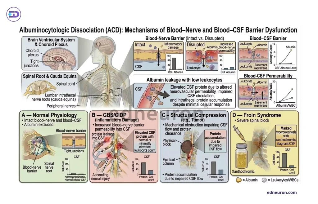
Fig 1 : Comparative panel describing the various mechanisms of ACD via disruption of the blood nerve barrier.
Horizon of Differentials
The differential diagnosis of albuminocytologic dissociation is broad enough to span nearly every subspecialty in medicine.
- Structural spine disease: herniated discs, spinal stenosis, spondylodiscitis, epidural lipomatosis- can produce modest protein elevation through chronic root compression.
- Leptomeningeal metastasis may present with ACD before malignant cells can be identifiable in the CSF, particularly early in the disease course, when cytology is falsely negative (36.4% of pooled patients had negative cytology, Palmisciano P et al. 2022)
- Neuroborreliosis, the neurological manifestation of Lyme disease, can mimic the electrophysiological (prolonged distal motor latency, conduction block, and absent F-wave response) and CSF profile of GBS sufficiently to mislead even experienced clinicians in endemic regions. This especially happens when the early presentation of Lyme’s disease, i.e., CSF pleocytosis, is absent.
- Amyloid neuropathy deserves particular mention: the diagnosis is frequently delayed because its pace, phenotype, and CSF findings overlap substantially with CIDP, and the clinical consequences of that delay – missed treatment for a progressive, multisystem disease are severe.
What unites these conditions is not a shared immunopathology but a shared consequential anatomical vulnerability: the blood–nerve and blood–CSF barriers, when disrupted by any sufficiently disruptive process, produce the same CSF signature. The signature is the clue. The diagnosis Must be rendered after much deliberation.
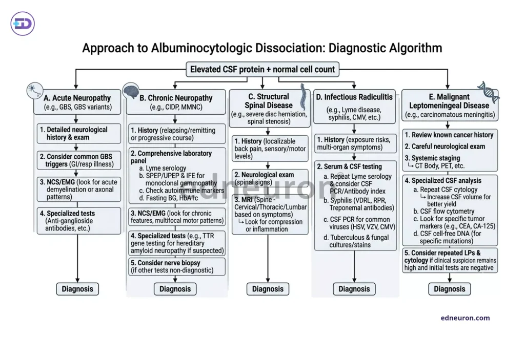
Fig 2 : Stepwise evaluation strategy.
Diagnostic Differentials: Reading the Clues
Three axes of reasoning are indispensable to reach the diagnosis:
- The clinical phenotype
- The temporal profile
- The pattern of ancillary findings.
Each etiology in the differential leaves a distinct imprint across these axes, and recognizing those imprints shall pave the path to the clinical endpoint.
- The temporal axis– the first discriminator:
- GBS is acute, and the nadir is typically reached within four weeks.
- CIDP, by definition, progresses beyond eight weeks, and its CSF protein often exceeds 100 mg/dL on serial sampling.
- Diabetic radiculoplexopathy follows a subacute course with spontaneous partial recovery – a trajectory that is incompatible with progressive GBS and should prompt metabolic re-evaluation.
- Structural spinal pathology, including Froin’s syndrome, tends to present with level-specific signs: a sensory level, upper motor neuron signs above the lesion, and sphincter dysfunction disproportionate to limb involvement.
- The phenotypic axis:
- Asymmetry is inconsistent with classical GBS and should raise suspicion for neuroborreliosis, vasculitic neuropathy, or structural compression.
- Cranial neuropathies accompanying radiculopathy in a patient from an endemic region demand Lyme serology before any other interpretation is accepted.
- Autonomic dysfunction: orthostatic hypotension, anhidrosis, gastrointestinal dysmotility that precedes or dominates the motor syndrome should immediately elevate amyloid neuropathy in the differential, even when CSF findings superficially resemble CIDP. Carpal tunnel syndrome and a family history of neuropathy are further amyloid flags that the CSF cannot provide.
- The ancillary investigation axis:
- Nerve conduction studies are not optional when ACD is found: the pattern of demyelination, its symmetry, the presence of conduction block, and the behavior of F-waves differentiate GBS subtypes, CIDP, and hereditary neuropathies with far greater specificity than protein elevation alone.
- MRI of the neuraxis should be obtained early if level-specific signs are present, if there is a known malignancy, or if the clinical course deviates from expectation.
- Leptomeningeal disease may be invisible on a single CSF cytology; repeat lumbar puncture and CSF flow cytometry substantially increase sensitivity when it is suspected.
- The CSF glucose, easily overlooked, may provide an early signal. Hypoglycorrhachia points toward infectious or malignant meningitis rather than immune neuropathy.
- Finally, in any patient with a neuropathy that resembles CIDP but responds incompletely, serum and urine electrophoresis, bone marrow biopsy, and TTR gene sequencing should be pursued before accepting the diagnosis of idiopathic CIDP as settled.
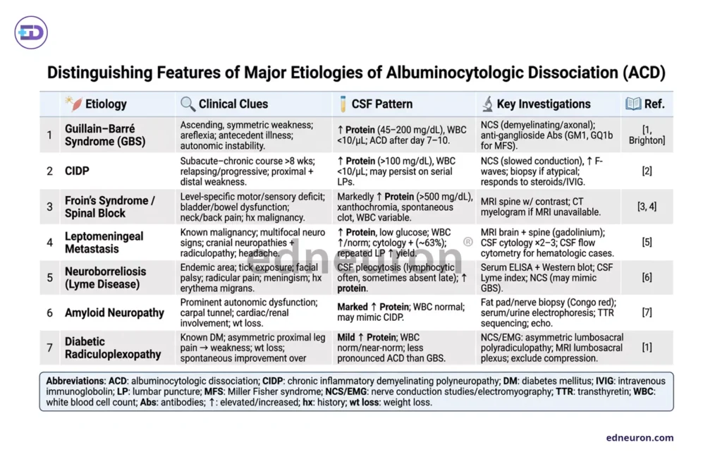
Fig 3 : A concise tabulated summary of all the clues leading to various differentials and their key investigations.
Conclusion
The original description by Guillain, Barré, and Strohl was an astute observational tendency rather than a rule, a probabilistic association between a CSF pattern and a clinical syndrome, observed in a limited number of carefully studied patients. The tendency is real. The rule was never proclaimed; it was transformed into one, unsolicited, for the sake of convenience when pattern recognition seems the easy way out, especially in high-volume, intense vigor clinical practice. Albuminocytologic dissociation, if properly understood, speaks to the clinician, indicating that the chemical barrier between blood and neural tissue has been disrupted. It does not specify the agent, the level, or the mechanism; rather, it points to a category of pathological processes. Within that category, the diagnostic range is broad enough to encompass immune neuropathy, spinal cord compression, malignant meningitis, and metabolic infiltration simultaneously.
The most important lesson of ACD is not what it confirms but what it cannot exclude. A clinician who encounters this finding and feels relieved has perhaps understood neurology as taxonomy rather than as reasoning. The more intellectually honest – and, ultimately, the more useful – response is a disciplined expansion of inquiry: imaging, nerve conduction studies, repeat CSF if necessary, and, always, a willingness to revisit the diagnosis when the patient fails to follow the expected course. In the end, the CSF report is a laboratory result. The diagnosis belongs to the clinician who holds the baton
References
- Sejvar JJ, Kohl KS, Gidudu J, Amato A et al. Brighton Collaboration GBS Working Group. Guillain-Barré syndrome and Fisher syndrome: case definitions and guidelines for collection, analysis, and presentation of immunization safety data. Vaccine. 2011 Jan 10;29(3):599-612. PMID: 20600491.
- Leonhard SE, Mandarakas MR, Gondim FAA, Bateman K, Ferreira MLB, Cornblath DR, et al. Diagnosis and management of Guillain–Barré syndrome in ten steps. Nat Rev Neurol. 2019;15(11):671-683.
- Van den Bergh PYK, et al. European Academy of Neurology/Peripheral Nerve Society guideline on diagnosis and treatment of chronic inflammatory demyelinating polyradiculoneuropathy: Report of a joint Task Force-Second revision. Eur J Neurol. 2021;28(11):3556–3583.
- Jacobs L, Delsaut B, Lamartine S, Monteiro M et al. Froin’s Syndrome: A Comprehensive Review of the Literature and the Addition of Two New Cases. Neurol Int. 2024 Sep 29;16(5):1112-1121. PMID: 39452685.
- Nouvakis D, Natsis KS, Tsika A et al. Progressive flaccid paraparesis with albuminocytologic dissociation: It’s not always Gullain-Barre syndrome. J Spinal Cord Med. 2020 Mar;43(2):276-278. PMID: 29939110.
- Palmisciano P, Sagoo NS, Kharbat AF et al. Leptomeningeal Metastases of the Spine: A Systematic Review. Anticancer Res. 2022 Feb;42(2):619-628. PMID: 35093859.
- Farr J, Bittar J. Neuroborreliosis Presenting as Guillain-Barré Syndrome. Cureus. 2023 Jul 23;15(7):e42322. PMID: 37614265.
- Li J, Li Y, Chen H et al. Autonomic Neuropathy and Albuminocytologic Dissociation in Cerebrospinal Fluid As the Presenting Features of Primary Amyloidosis: A Case Report. Front Neurol. 2017 Jul 26;8:368. PMID: 28798722.

Author: A P
A medical trainee with an emerging focus on translational and clinical research, with interests spanning surgical sciences, neuroscience, pediatrics, and immunology. Her academic trajectory reflects an effort to integrate molecular innovation with clinically relevant disease models, particularly in complex and high-burden conditions. Her research experience includes work in genome engineering, specifically in prime editing, exploring its therapeutic potential in precision medicine. She has also contributed to oncological research examining cholangiocarcinoma with brain metastasis, focusing on its clinical course and diagnostic challenges. In parallel, her work investigating stoma formation as an independent risk factor for acute kidney injury reflects an interest in perioperative and systemic complications. Academically, she has contributed to case-based and review-driven scholarship, including a case reports and interdisciplinary review articles. Her evolving interests in neurology, pediatrics, and immunology reflect a broader inclination toward understanding disease across systems—from molecular mechanisms to clinical outcomes—while maintaining a disciplined, evidence-based approach to patient care. MBBS (MS4) ABVIMS Dr. RML HOSPITAL New Delhi